
An electrocardiogram records the electrical signals in your heart. It’s a common and painless test used to quickly detect heart problems and monitor your heart’s health.
Electrocardiograms — also called ECGs or EKGs — are often done in a doctor’s office, a clinic or a hospital room. ECG machines are standard equipment in operating rooms and ambulances. Some personal devices, such as smart watches, offer ECG monitoring. Ask your doctor if this is an option for you.
Why it’s done
An electrocardiogram is a painless, non-invasive way to help diagnose many common heart problems in people of all ages.
Your doctor may use an electrocardiogram to determine or detect:
Abnormal heart rhythm (arrhythmias)
If blocked or narrowed arteries in your heart (coronary artery disease) are causing chest pain or a heart attack
Whether you have had a previous heart attack
How well certain heart disease treatments, such as a pacemaker, are working
You may need an ECG if you have any of the following signs and symptoms:
Chest pain
Dizziness, lightheadedness or confusion
Heart palpitations
Rapid pulse
Shortness of breath
Weakness, fatigue, or a decline inability to exercise.
Risks
An electrocardiogram is a safe procedure. There is no risk of electrical shock during the test because the electrodes used do not produce electricity. The electrodes only record the electrical activity of your heart.
You may have minor discomfort, similar to removing a bandage when the electrodes are removed. Some people develop a slight rash where the patches were placed.
How you prepare
No special preparations are necessary for a standard electrocardiogram. Tell your doctor about any medications and supplements you take. These can often affect the results of your test.
What you can expect
An electrocardiogram can be done in a doctor’s office or hospital and is often done by a nurse or technician.
Before
You may be asked to change into a hospital gown. If you have hair on the parts of your body where the electrodes will be placed, the technician may shave the hair so that the patches stick.
Once you’re ready, you’ll be asked to lie on an examining table or bed.
During
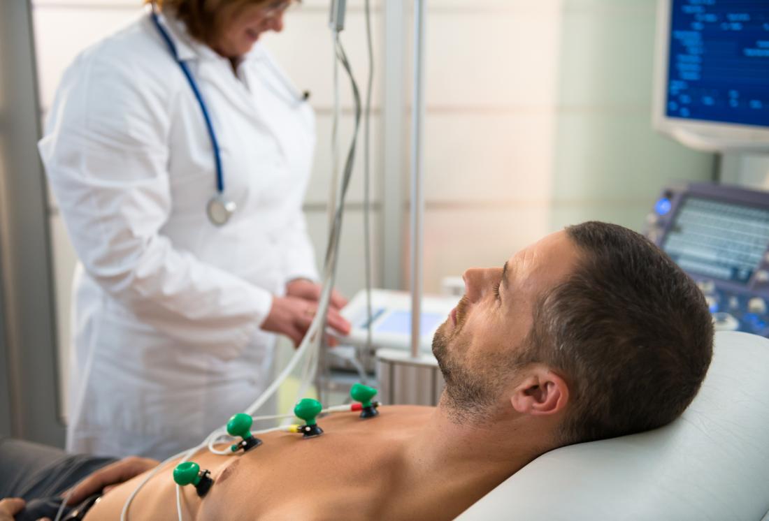
During an ECG, up to 12 sensors (electrodes) will be attached to your chest and limbs. The electrodes are sticky patches with wires that connect to a monitor. They record the electrical signals that make your heartbeat. A computer records the information and displays it as waves on a monitor or on paper.
You can breathe normally during the test, but you will need to lie still. Make sure you’re warm and ready to lie still. Moving, talking or shivering may distort the test results. A standard ECG takes a few minutes.
After
You can resume your normal activities after your electrocardiogram.
Results
Your doctor may discuss your results with you the same day as your electrocardiogram or at your next appointment.
If your electrocardiogram is normal, you may not need any other tests. If the results show an abnormality with your heart, you may need another ECG or other diagnostic tests, such as an echocardiogram. Treatment depends on what’s causing your signs and symptoms.
Your doctor will review the information recorded by the ECG machine and look for any problems with your heart, including:
Heart rate. Normally, heart rate can be measured by checking your pulse. An ECG may be helpful if your pulse is difficult to feel or too fast or too irregular to count accurately. An ECG can help your doctor identify an unusually fast heart rate (tachycardia) or an unusually slow heart rate (bradycardia).
Heart rhythm. An ECG can show heart rhythm irregularities (arrhythmias). These conditions may occur when any part of the heart’s electrical system malfunctions. In other cases, medications, such as beta-blockers, cocaine, amphetamines, and over-the-counter cold and allergy drugs, can trigger arrhythmias.
Heart attack. An ECG can show evidence of a previous heart attack or one that’s in progress. The patterns on the ECG may indicate which part of your heart has been damaged, as well as the extent of the damage.
Inadequate blood and oxygen supply to the heart. An ECG done while you’re having symptoms can help your doctor determine whether chest pain is caused by reduced blood flow to the heart muscle, such as with the chest pain of unstable angina.
Structural abnormalities. An ECG can provide clues about enlargement of the chambers or walls of the heart, heart defects and other heart problems.
If your doctor finds any problems on your ECG, he or she may order additional tests to see if treatment is necessary.